A discovery at SR-Tiget reveals the key liver cells driving organ growth and advancing pediatric gene therapy
Researchers at the San Raffaele Telethon Institute for Gene Therapy (SR-Tiget) have discovered that only a minority of cells in the newborn liver are responsible for generating most of the adult organ. The study, published in the Journal of Hepatology, sheds new light on liver development and offers valuable insights to design more effective gene therapies for children with genetic liver diseases.
The team showed that just 15–20% of hepatocytes — the main type of liver cell — in newborn mice, called clonogenic hepatocytes, give rise to more than 90% of the adult liver.
Understanding how these cells grow and mature early in life is crucial to improve strategies that aim to correct inherited diseases directly inside the liver, such as gene transfer or genome editing.
Cutting-Edge Tools to Map Liver Development
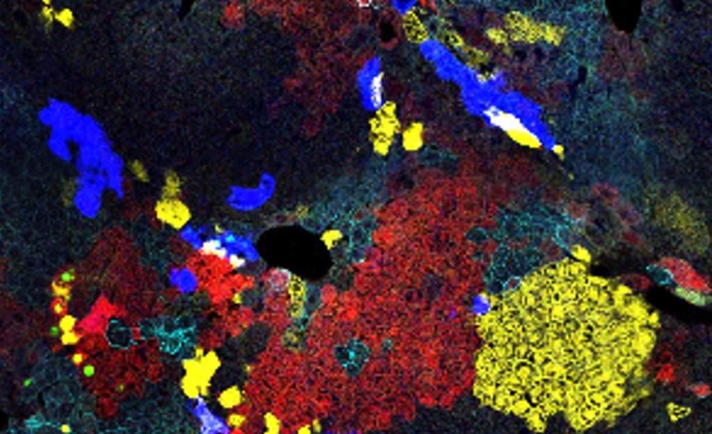
To reach these results, researchers combined some of the most advanced techniques available today — including single-cell and spatial transcriptomics, clonal tracing and mathematical modeling. This allowed them not only to identify the clonogenic hepatocytes but also to understand the signals and microenvironments that regulate their activity.
“By mapping the exact location and identity of these cells, we gained an unprecedented view of how the liver grows after birth,” explains Dr. Michela Milani, co-first author of the study.
Towards More Effective Pediatric Therapies
The findings also show that the success of gene therapy approaches depends on both the timing of treatment and the identity of the targeted cells. For instance, certain types of gene editing work particularly well in clonogenic hepatocytes, making their corrected genetic information spread more widely as the liver grows.
Other approaches, like the use of viral vectors, are influenced by how the liver matures and by specific zones of the tissue that become less receptive to gene transfer over time.
“Knowing which cells to target — and when — is essential to make gene therapies more durable,” says Dr. Francesco Starinieri, co-first author.
The study also revealed that clonogenic hepatocytes in newborn livers are located next to blood progenitor cells, suggesting that they share growth signals. This discovery could open up new opportunities for regenerative medicine.
“This research brings us closer to developing more effective and long-lasting gene therapies for children”
Alessio Cantore
“By identifying the specific cells that fuel liver growth, we can design treatments that work better and last longer” comments Dr. Alessio Cantore, senior and corresponding author.
A collaborative project
The work was carried out in collaboration with Dr. Andrés Muro of the International Centre for Genetic Engineering and Biotechnology (ICGEB) in Trieste, with contributions from biologists, physicists and bioinformaticians at SR-Tiget and the San Raffaele Scientific Institute.
The study was supported by Fondazione Telethon, the Italian Ministry of Health, and the European Union’s Horizon 2020 program. Dr. Cantore also recently received a Consolidator Grant from the European Research Council (ERC) to continue investigating liver development and its therapeutic applications.