The TIGEM LEAD-ER project investigates whether ER-phagy can be pharmacologically activated to remove misfolded proteins involved in different diseases.
There is a pivotal moment in the scientific process: the point at which research originally undertaken to understand how a biological process works begins to be explored from an applied perspective, to determine whether it might eventually provide the basis for a therapeutic solution.
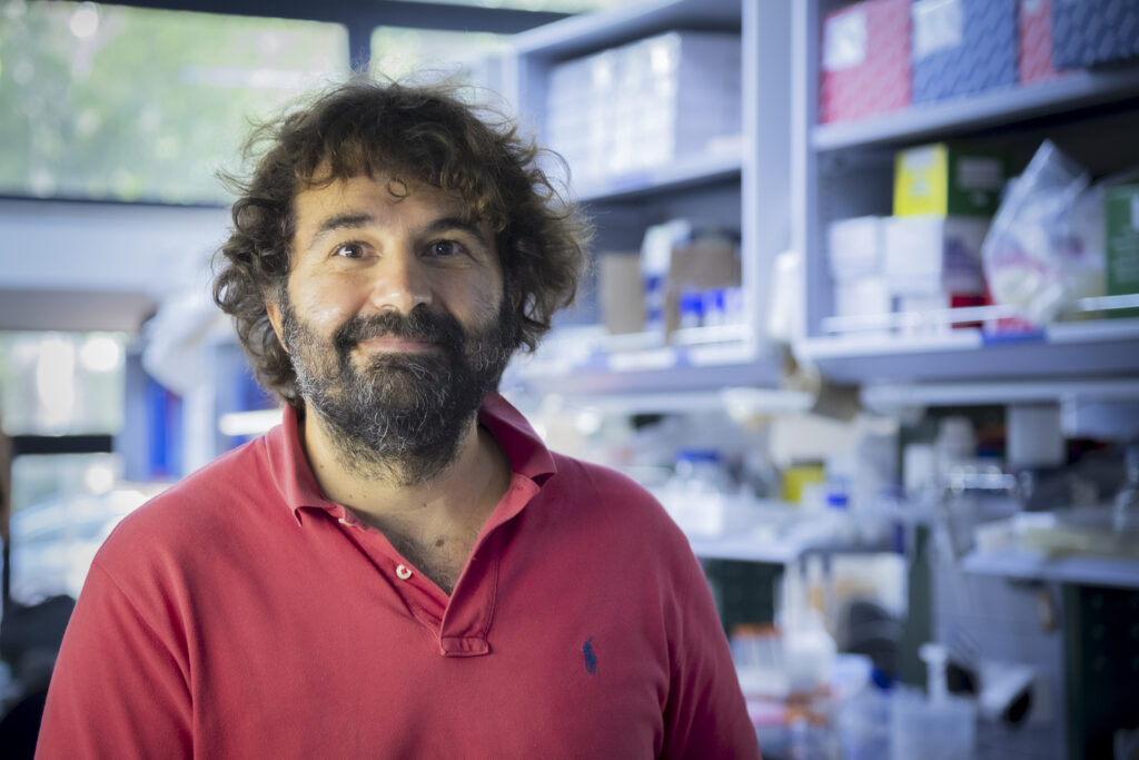
This is what is happening with LEAD-ER, the ERC Proof of Concept grant awarded to Carmine Settembre, Professor of Histology at the University of Naples Federico II and Principal Investigator at TIGEM. Announced on 30 June, the grant focuses on the translational development of ER-phagy, a form of selective autophagy that enables cells to remove portions of the endoplasmic reticulum together with damaged proteins that might otherwise accumulate, form aggregates and become toxic. The project does not aim to activate autophagy indiscriminately, but rather to determine whether it is possible to target the receptors that make this process selective.
LEAD-ER builds on AUTO-SELECT, Settembre’s previous ERC Consolidator Grant, through which he investigated selective autophagy at the molecular level. The project therefore marks a transition from understanding the process to exploring the possibility of intervening in it. “Having demonstrated how this mechanism works, with the new funding we now want to understand whether we can modulate it pharmacologically” Settembre explains.
This important research is reshaping our understanding of protein degradation systems and drawing attention to a cellular quality-control mechanism that could be relevant to diseases characterised by the accumulation of defective proteins within the endoplasmic reticulum.
ER-phagy: selective autophagy of the endoplasmic reticulum
To understand why ER-phagy represents such an important development, it is necessary to start with autophagy: the process through which cells degrade and recycle damaged or no longer functional intracellular components. This mechanism is essential for maintaining cellular homeostasis, particularly when altered proteins or impaired organelles begin to accumulate and interfere with normal cellular function.
For a long time, however, autophagy was regarded as a relatively non-selective disposal system. It was known that cells could degrade parts of themselves, but it was less clear how they were able to determine precisely what should be removed. In recent years, this picture has changed with the identification of autophagy receptors: proteins capable of recognising specific substrates and promoting their incorporation into autophagosomes, vesicles that transport them to lysosomes for degradation.
This is the context in which TIGEM’s work on autophagy of the endoplasmic reticulum is situated.
The endoplasmic reticulum is involved in the production and folding of a large proportion of cellular proteins. This makes it an extremely important, but also particularly vulnerable, cellular compartment. Protein production is inherently prone to error: some of the proteins produced fail to adopt the correct conformation and must therefore be eliminated.
For many years, the management of misfolded proteins within the endoplasmic reticulum was understood primarily in terms of the unfolded protein response and degradation systems associated with the proteasome. TIGEM’s research has shown, however, that this is not the only mechanism available.
Alongside these systems, cells also rely on ER-phagy, a selective form of autophagy that enables them to remove not only individual proteins, but entire portions of the endoplasmic reticulum in which misfolded or aggregated proteins have accumulated, before they become toxic. ER-phagy therefore acts as a genuine quality-control mechanism for the endoplasmic reticulum.
The process is regulated by specific ER-phagy receptors. On the luminal side of the endoplasmic reticulum, these receptors interact with molecular chaperones associated with misfolded proteins, helping to identify the cargo that must be removed. On the cytoplasmic side, the same receptors recruit the autophagy machinery. This is the step that allows the cell to dispose specifically of the portion of the endoplasmic reticulum identified as damaged.
The result is a much more precise system than the generalised activation of autophagy. The cell does not need to degrade mitochondria, ribosomes or cytoplasmic proteins indiscriminately. Instead, it must recognise a specific problem located within the endoplasmic reticulum and direct the degradation process towards that precise site.
This is why TIGEM’s research has focused on ER-phagy receptors. These receptors make the selectivity of the process possible by binding misfolded proteins within the endoplasmic reticulum on one side, while recruiting the autophagic machinery required to eliminate them on the other.
A single potential pharmacological target for different diseases
Even under normal conditions, a small proportion of the proteins produced by a cell fail to fold correctly and must be eliminated. This is a physiological margin of error that cellular quality-control systems routinely manage. When a mutation alters a protein, however, this proportion can rise to almost 100%, disrupting the balance. “The amount of protein that begins to accumulate in the endoplasmic reticulum exceeds the cell’s capacity to remove and degrade it” Settembre explains.
This is precisely the imbalance that the project aims to correct by increasing the removal of defective proteins accumulated within the endoplasmic reticulum. “By increasing the rate of degradation, we are trying to slow down the rate at which these proteins are deposited and form aggregates” Settembre continues.
The same problem recurs, in almost identical form, across diseases that may appear to have nothing in common. In many of these conditions, the damage is caused not only by the loss of a functional protein, but also by the toxicity of its mutated form, which remains trapped within the endoplasmic reticulum. This is the case in several autosomal dominant disorders, in which one copy of the gene produces a normal protein while the other produces an altered form.
The same underlying mechanism is found in a range of diseases, including alpha-1 antitrypsin deficiency, which affects the liver and lungs; osteogenesis imperfecta, which affects the skeleton; retinitis pigmentosa, which involves the eye; certain forms of diabetes; and uromodulin-associated disease, a kidney disorder. Different organs, different symptoms, but the same cellular defect. In retinitis pigmentosa, for example, photoreceptor death is not caused simply by a lack of rhodopsin, but by endoplasmic reticulum stress resulting from the accumulation of the misfolded protein.
It is precisely this cross-cutting relevance that makes the project potentially significant beyond individual rare diseases. If the mechanism is shared, the same family of molecules capable of promoting the degradation of these proteins could, in theory, have applications across several conditions. The broader perspective also includes more common conditions, such as ageing and obesity, in which endoplasmic reticulum stress plays an important role.
Turning ER-phagy into a pharmacological target
The question now is whether this mechanism can be controlled from outside the cell. To turn ER-phagy into a potential therapeutic target, it must be activated in a controlled, selective and measurable way.
The strategy at the heart of the project is to identify small molecules capable of activating ER-phagy receptors. “The target for intervention is the cytosolic component of the receptors, which recruits the autophagy proteins and enables the formation of the autophagosome” Settembre explains.
The assay developed in the laboratory measures receptor activation through an indirect signal: its association with the proteins involved in autophagosome formation. If a molecule increases this association, it can be selected as a candidate because it may have promoted the recruitment of the autophagic machinery and initiated the degradation of the affected portion of the endoplasmic reticulum.
This makes it possible to carry out high-throughput screening and test a very large number of compounds. The most promising molecules are then selected and subjected to further assays, with the aim of identifying a family of compounds that is active, at least in vitro. The next step is validation in cells: determining whether, in the presence of the molecule, the degradation of mutated proteins accumulated within the endoplasmic reticulum is effectively accelerated.
Alongside pharmacological screening, the laboratory also uses a genetic validation approach. Before searching for the right molecule, it is necessary to demonstrate that enhancing ER-phagy is beneficial in the disease in question.
To do this, the receptors are overexpressed in cellular models. If the accumulation of defective proteins decreases, or if the cellular phenotype improves, this indicates that the disease may respond to ER-phagy activation.
This helps determine whether ER-phagy is a valid target. If the target is confirmed, the failure of a particular molecule would not rule out the mechanism itself, but would instead indicate that a better compound needs to be identified or optimised.
The next stage will involve chemical optimisation. The most promising compounds can be modified to improve their specificity, stability and biodistribution, while reducing toxicity and off-target effects.
ER-phagy as a pharmacological target: the challenges ahead
Alongside the pharmacological screening, however, a fundamental question remains open: what activates ER-phagy receptors?
The receptor must not only be present on the membrane of the endoplasmic reticulum, but must also become capable of recruiting the autophagic machinery and initiating autophagosome formation. The difficulty arises from the same structural feature that also makes pharmacological development challenging: the region of the receptor involved in this step is intrinsically disordered and lacks an easily predictable structure. This makes it difficult both to understand how it is activated and to design molecules capable of binding to it effectively.
Another question is whether activating endogenous receptors would be sufficient to produce a therapeutic effect. “We are still at the hypothesis stage,” Settembre explains. “We do not yet know whether activating the endogenous levels of these proteins can achieve the same degree of therapeutic relevance”.
A potentially favourable factor may, however, lie in the biology of the system itself. Under normal conditions, the receptors are expressed at low levels, whereas their expression increases in cells experiencing endoplasmic reticulum stress. “This could increase the specificity of the drug, as it would target a protein that is expressed at very low levels in healthy cells and at higher levels in diseased cells” Settembre comments.
Safety is another important challenge. Activating a cellular degradation system requires a balance to be found between efficacy and the risk of interfering with other cellular processes. From this perspective, the pharmacological approach offers a significant advantage, because the dose and activity of a molecule can be adjusted and controlled.
At this stage, the aim is not to promise an imminent therapy, but to determine whether a mechanism shared by different diseases — the accumulation of defective proteins within a stressed endoplasmic reticulum — can become a pharmacologically modulatable target. If this hypothesis is confirmed by TIGEM laboratories, ER-phagy could offer a new way of addressing different conditions, starting not from an individual gene or organ, but from a shared defect in cellular quality control.
This is precisely the strength of the approach: not to replace the cell’s quality-control system, but to determine whether it is possible to promote the removal of misfolded proteins when the endoplasmic reticulum is under stress. This is how a discovery in cell biology can begin to become a potential selective and controllable pharmacological strategy.
